When first using EMG it can be difficult to identify a “good” emg recording from a “bad” one. Here are some things you should look for:
- Distinct base lines
- Distinct muscle activation
- Signals zeroed about zero volts with almost a mirror between the positive and negative chart areas
- Comparable signals between left and right muscles with similar voltages and activation times (may not be applicable with some populations)
- Comparable voltage levels across all muscle recordings (may not be applicable with some populations)
Below are some examples that may be helpful when diagnosing your signal.
A “Good Signal”
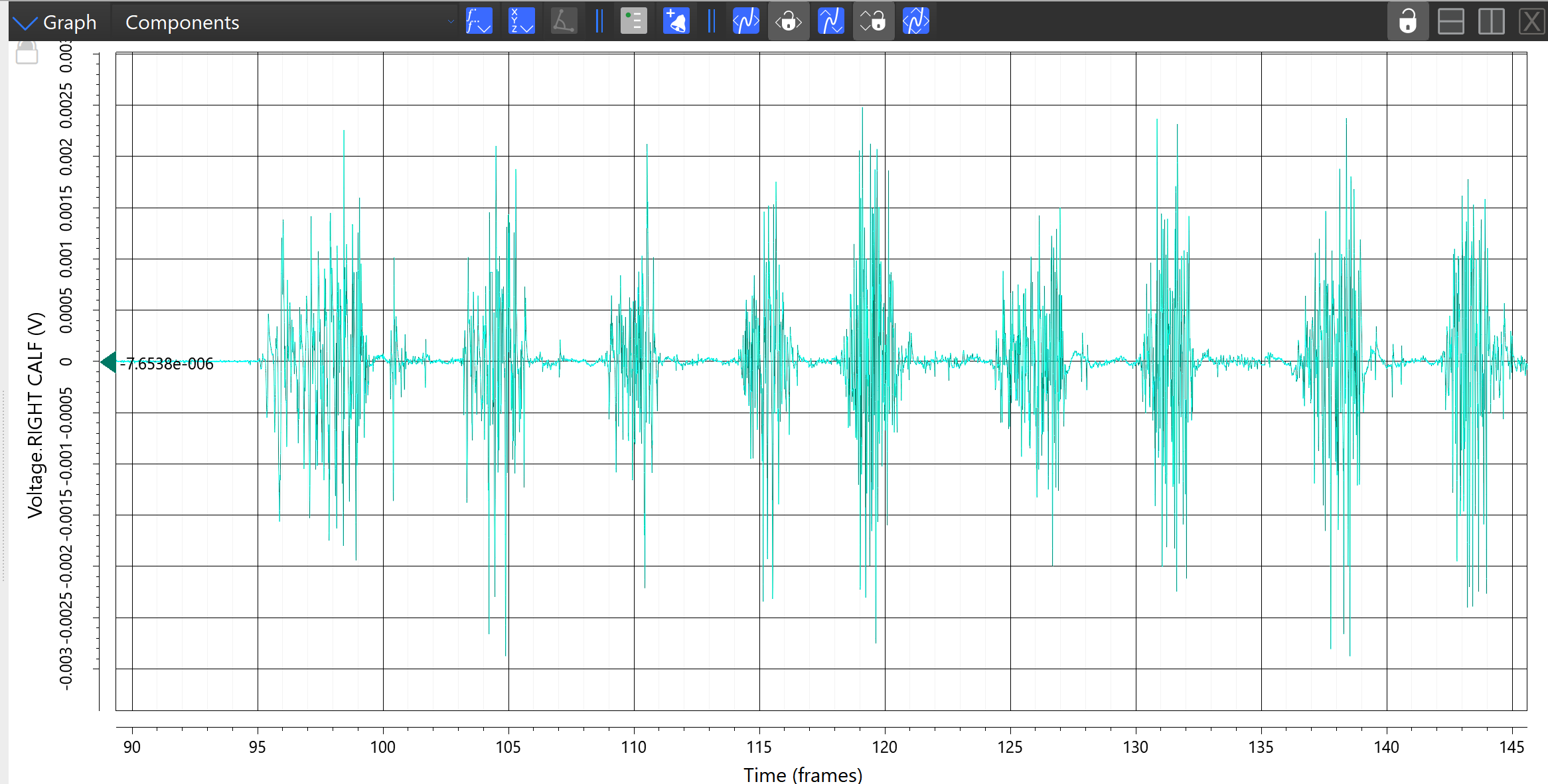
Here is an example of a fairly good raw EMG signal. Take not of the regular voltage bursts with clear breaks in between.

A Noisy Signal
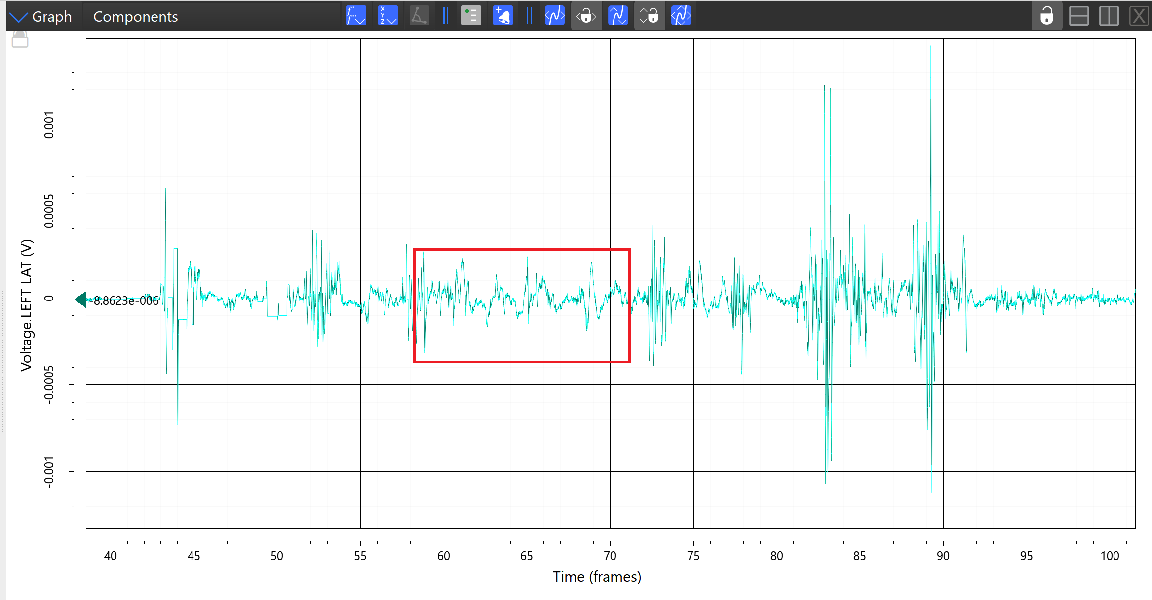
The below signal is somewhat noisy. The area outlined in red is of particular concern due to it’s irregular shape. This may be movement artefacts interrupting the signal.

Signal Cutout
At the 85 second mark of the signal below the signal is dropped and does not reconnect until around the 95 second mark. This almost looks like the signal has flat-lined, even at rest a a signal will not look unnaturally flat or be so far offset from the zero line.

ECG artefact
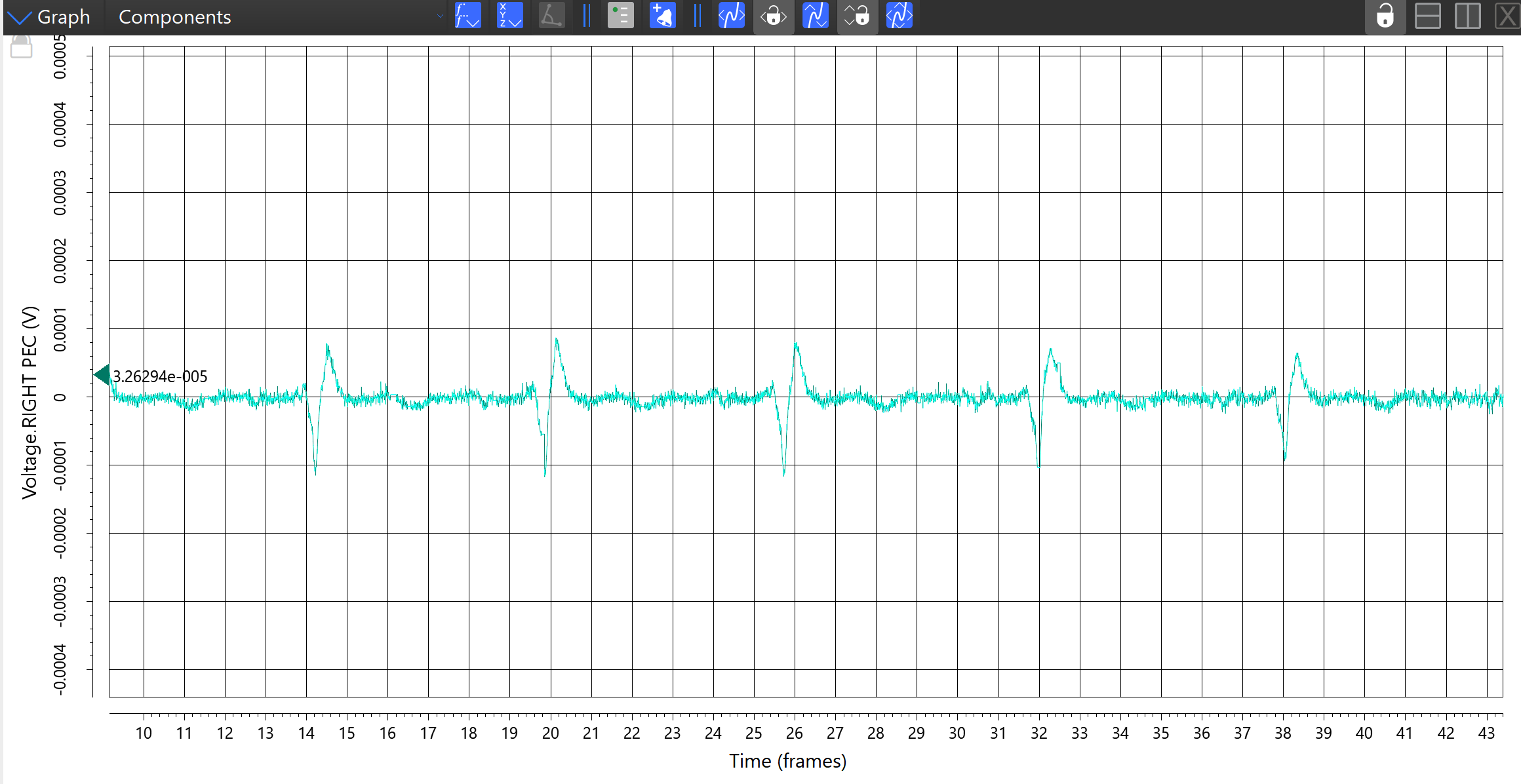
The following EMG was taken from a pectoral muscle during a recording. Notice the irregular shaped bump occurring at a regular 6 second interval? This is from the heart muscles.

There are of course many more kinds of signal noise and interference, this guide is only intended to provide some examples to help identify if there is an obvious issue with your signal.
Having trouble with signal disconnections? We have a guide here that you might find helpful.

how can i convert my ADC results in mV in collecting EMG signal?
I am Using AD8232 chip